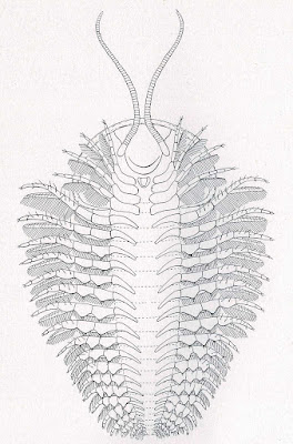
Yesterday's posting was of the trilobite Triarthrus eatoni (Hall, 1838) specimens on display at the Harvard Museum of Natural History. Today's post will cover another species of this trilobite highlighted in The Appendages, Anatomy, and Relationships of Trilobites by Percy E Raymond. It was published in December of 1920 by Connecticut Academy of Arts and Sciences. Dr. Raymond was associate professor of palaeontology and curator of invertebrate palaeontology in the Museum of Comparative Zoology at Harvard University. It covers Charles Emerson Beecher (1856-1904) unpublished work on trilobites including the Triarthrus becki (Green, 1839?) found in the Utica Shale. Paleo-illustrator and MIT/Columbia University educated paleontologist Elvira Wood (1865-1928) did a lot of illustrating on this book.
On page 40 a historical description was provided, "Specimens of Triarthrus retaining appendages were first obtained by Mr. W. S. Valiant from the dark carbonaceous Utica shale near Rome, New York, in 1884, but no considerable amount of material was found until 1892. The first specimens were sent to Columbia University, and were described by Doctor W. D. Matthew (1893). This article was
accompanied by a plate of sketches, showing for the first time the presence of antennules in trilobites and indicating something of the endopodites and exopodites of the appendages of the cephalon, thorax,
and pygidium. Specimens had not yet been cleaned from the lower side, so that no great amount could then be learned of the detailed structure. Matthew concluded that 'The homology with Limulus seems
not to be as close in Triarthrus as in the forms studied by Mr. Walcott; but the characters seem to be of a more comprehensive type, approaching the general structure of the other Crustacea rather than any special form.'"
The following are from plate I in the book. Described as "Photographs of Triarthrus becki, made by C. E. Beecher."
Fig. 1. Specimen 213. The dorsal test has been removed from the glabella, revealing the outline of the posterior end of the hypostoma, the proximal ends of the antennules, the gnathites, and incomplete
endopodites of some appendages, × 5.43
right side, × 7.33.
Fig. 3. Specimen 217. This specimen shows better than any other the form of the gnathites of the cephalon. Note also the setæ of the exopodites under the cheek at the right. The appearance of a hook on the posterior gnathite on the right may be accidental, but it does not show broken edges, × 6.85.
Fig. 4. Specimen 215. The ventral side of the cephalon of a small entire specimen. Shows well the form of some of the gnathites and a few of the endopodites. Note the unusual position of the antennules. × 7.63.
Fig. 5. Specimen 226. This specimen did not photograph well, but is important as showing the exopodites and endopodites emerging from under the cephalon. × about 6.


No comments:
Post a Comment