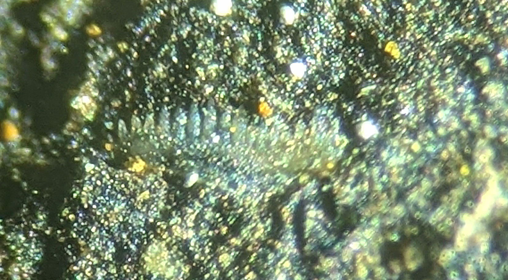
Here are two freshly found conodont fossils in the New Albany Shale (Late Devonian Period). The first appears to be some sort of Spathognathodus sp. (Branson and Mehl, 1941). The second appears to be of the genus Elictognathus (Cooper, 1939). Both were found on a shale plate about 4 cm by 4 cm. Field of view for images was about 1 mm. Specimens were collected in Floyd County, Indiana, USA. Indiana Geological Survey Bulletin 41, Conodonts from the Jacob Chapel Bed (Mississippian) of the New Albany Shale in Southern Indiana by Carl B. Rexford (1969) was used for the identification of these fossils.
This fossil insect was found in the Florissant Formation
of Teller County, Colorado USA. It appears to be march fly. Fossil
dates to the Eocene Epoch of the Paleogene Period (about 34 million
years ago). Field of views is approximately 7 mm.Thanks to Kenny for the image.
This fossil insect was found in the Florissant Formation of Teller County, Colorado USA. It appears to be midge fly. Fossil dates to the Eocene Epoch of the Paleogene Period (about 34 million years ago). Field of views is approximately 5 mm.
The image shows a snout beetle or weevil insect fossil of the Order
Coleoptera, Family Curculionidae found in the Florissant Formation of
Teller County, Colorado, USA. It dates to the Eocene Epoch of the
Paleogene Period. Thanks to Doug for the image.
This image shows an ant insect fossil found in the Florissant Formation of Teller County, Colorado, USA. The field of view (FOV) is 5 mm. It dates to the Eocene Epoch of the Paleogene Period.
These images show unidentified fossils found in the
Florissant Formation of Teller County, Colorado, USA. The field of view
(FOV) for the top one is 2 mm and it could be a fragment of horsetail plant with predation holes. The second could be a seed or a coprolite and its FOV is 5mm. They date to the Eocene Epoch of the Paleogene Period.
This image shows an unidentified insect larva fossil found in the Florissant Formation of Teller County, Colorado, USA. The field of view (FOV) is 3 mm. It dates to the Eocene Epoch of the Paleogene Period.
This microfossil appears to be a Chiracanthus sp. fish scale.
The field of view (FOV) is about 2 mm. It dates to the Devonian Period. The fossil was found in the Beechwood
Limestone of
Clark County, Indiana USA.
This image shows what appears to be a Polygnathus longiposticus conodont fossil. This creature existed in the Mississippian Period. Fossil was found in the Jacob Chapel Shale of Clark County Indiana USA.
Source for ID is Conodonts of the Jacobs Chapel Shale by Carl B. Rexroad, Indiana Department of Natural Resources Geological Survey Bulletin 41 (published Bloomington Indiana 1969).
This image shows what appears to be a Spathognathodus sp. conodont fossil. This creature existed in the Mississippian Period. Fossil was found in the Jacob Chapel Shale of Clark County Indiana USA.
Source for ID is Conodonts of the Jacobs Chapel Shale by Carl B. Rexroad, Indiana Department of Natural Resources Geological Survey Bulletin 41 (published Bloomington Indiana 1969).
This image is of a microfossil fragment that appears to be an Atraktoprion scolecodont. It was found in the Kope or Fairview Formations of Mason County, Kentucky, USA. The creature existed in the Ordovician Period. Field of view is about 1 mm.
This fossil appear to be Polygnathus cristatus? conodont found in the Middle Devonian Beechwood
Limestone of Clark County, Indiana. Helicon Focus was used to create some of these images. This fossil is amber colored and translucent so a underneath light was used to reveal more detail.
These fossils appear to be Ozarkodina willsi? conodonts found in either the upper Devonian New Albany Shale or middle Devonian Beechwood Limestone of Clark County, Indiana.
See the conodont.info web site to see Ozarkodina polita specimens: CLICK HERE and Ozarkodina tenuis specimens: CLICK HERE
A friend sent me an e-mail the other day with a link to the Indiana9fossils website microfossil section. I looked at those prices and said "YIKES!"
It shows a number of Devonian period condonts from the Vernon Limestone of Indiana. Notice the pictures of the Hibbarella sp and Polygnathus cristatuscondonts and the compare them to the pictures on D&D Fossil site in the United Kingdom. Well I do declare, it looks like the picture labeled Polygnathus from the New Albany Shale is the same. The condont labeled Hibbarella has the same picture as well. Hope everybody is cool with the copyrights on these images since both are commercial ventures.
The fossils on the Indiana9fossil site are being sold at $10 a piece while D&D Fossil is selling a capsule to small bag of mixed fossils for $10. So it appears if you want to save money buy the container and use the nice ID card to determine what fossils you have.
There appears to be some difference as to what layer these fossils are from while one site says Vernon Limestone and the other New Albany Shale but the file name shows Beechwood. It looks like the ID sheet shows them from either New Albany Shale or Beechwood Limestone. Speed, Indiana is a quarry/concrete plant so it would be interesting who provided this material from the quarry and then who dissolved it in acid to get extract the fossils. I have not e-mailed anyone at either dealer site to ask any questions. This set of fossils origin make for a good mystery. Delve some more into to the microfossil sections of both sites and more overlapping fossils will be found.
The net margin is pretty good if a bag yields 10-20 fossils that can be sold for $10 a piece. Of course, they need to be identified, packaged and shipped.
Yes, you read the title correctly. ASPEX Corporation of Delmont, Pennsylvania is offering the public the chance to have an item imaged with their Scanning Electron Microscope (SEM).
Image compliments of ASPEX Corporation
According to their literature, an optical microscope achieves a usable image at magnification of 100 times and can go up to 1000 times (20 micrometers). The ASPEX Personal Scanning Electron Microscope can image an object to 25 nanometers (nm) and has a scanning range of 100 nm to 5 mm. They go on to stress the resolution is not the only variable to consider when analyzing a specimen. SEMs provide a more depth-of-field than optical microscopes.
There are a wide range of applications SEMs can be used for and not just in the research laboratory. ASPEX makes an application for forensics (e.g. like a real world CSI) specifically Gunshot Residue (GSR) analysis. Other applications that can take advantage of SEM analysis: industrial production quality control, metal quality measurements, aerospace/defense maintenance prediction, and the pharmaceutical production.
All and all this is a great opportunity for amateur paleontologists. I keep wondering what items would I like to see: foraminifera, scolecodont tooth, ridge in cornulite tube, small fluorite crystals, pyrite crystal, growth lines on modern snail shell, crinoid stem segment or a pore of a bryozoan. So many possibilities in the microscopic world to explore!
One needs to remember that unlike optical microscopes, SEMs image in grayscale so no color output.
Well, I encourage those interested in exploring this generous offer made by ASPEX Corporation and check out the images already displayed at their website.
How to Participate in the "Send Us Your Sample" Campaign
1. Download and print this form from the ASPEX website.
2. Fill out the form and mail it along with the sample to be scanned:
ASPEX Corporation Free Sample Submissions 175 Sheffield Dr. Delmont, PA 15626
3. Once ASPEX has completed the scan, the images and report will be posted on ASPEX's website here.
Note: It should take about 2 weeks [UPDATE: 05/17/2010 I received the results from the fossil of an Ordovician Period worm tube. See the results at this post.] for the results to post to the ASPEX website, and the submitters will be notified via e-mail. If you want your sample returned, please say so on the submission form.
Remember to follow common sense on sending material to be scanned and also follow all postal regulations for shipping specimens through the mail.
These microfossils were found in the Lexington Limestone of Franklin County, Kentucky. They existed in the Ordovician Period. This first fossil is a snail or gastropod. It appears to be composed of some sort of black material. All fossils are about 1 mm in length. A microscope was used to magnify 40x.
I tried something new to help produce an in focused image of these 3-D fossils when using a microscope. The idea was to take a series of images with the microscope of one part of the fossil in focus. After taking a series of 4-6 images, put them in a program like Registax to stack the in focus parts together. Well, the plan hit some snags due to my process. First, I was using a handheld camera so the fossil was in slightly different place in each file. The fossil was too big for target box to follow in Registax. The program was designed to be used with astronomy images taken with the telescope so a 16x16 or 64x64 pixel area can be tracked when it is planet or crater size in the image. Needless to say the stacked image was some what blurry.
I then tried using Photoshop to align and stack the images. The program was geared to work with larger images. The microfossils images were cropped to about 700x700 pixels and were aligned. I then realized that even though the microscope had been set to focus on a different area of the 3-D fossil the camera I was using seemed to being focusing on the same area of the fossil each time. So all my images pretty much look the same thus rendering the stacking ineffective! I will try this experiment again at a later date and with a fixed mounted microscope camera.
This fossil might be a crinoid stem fragment.
Thanks to an identification by blog visitor Solius. This is a "Hyolithos; an enigmatic creature that some propose are mollusks."
These microfossils were found in the Lexington Limestone in Franklin County, Kentucky. They appear to be some sort of spines maybe belonging to a crinoid. Magnification was set to 40x. Fossils are from the Ordovician Period.